Veterinary advice should be sought from your local veterinarian before applying any treatment or vaccine. Not sure who to use? Look up veterinarians who specialize in poultry using our directory listing. Find me a Vet

Sarcocystosis

Rice Breast Disease

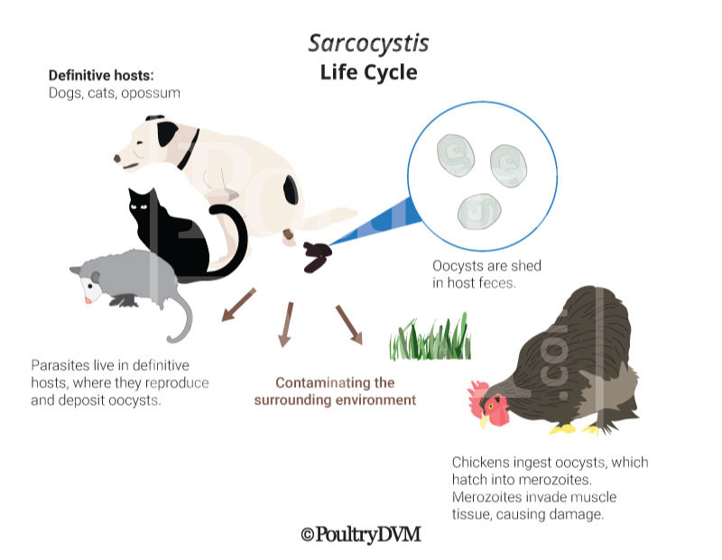
Sarcocystosis is an infection caused by a protozoan parasite of the genus Sarcocystis, which includes more than 200 known species. Ducks are affected by S. horvathi and S. wenzeli, which are found in domestic dogs and cats.

Ducks become infected with the parasite by ingesting Sarcocystis oocysts (eggs) shed in dog or cat feces.
Oocysts can remain infective in the environment for months. Once ingested, oocysts hatch into merozoites. Merozoites invade the duck's muscle tissue, causing damage.

Sarcocystosis can be diagnosed by identifying the cysts within muscle tissue or by identifying oocysts in the feces during a fecal test.

Symptoms

Torticollis (wry neck)
Paralysis
Trembling
Polyuria
Depression

Diagnosis

  • History
  • Clinical signs
  • Physical exam
  • Necropsy

Treatment

MethodMethod Summary
Supportive careIsolate the bird from the flock and place in a safe, comfortable, warm location (your own duck "intensive care unit") with easy access to water and food. Limit stress. Call your veterinarian.
Pyrimethamine
Trimethoprim-sulfadiazine60 mg/kg PO, SC q12h x 3 days, off 2 days, on 3 days

Reported Cases

  • Case 1: Encephaltiis sarcocystisis in a Psittacines A total of 5 psittacine birds in an enclosed zoological exhibit, including 2 princess parrots and 3 cockatoos of 2 different species, developed severe central nervous system clinical signs over a 2-3-month period and died or were euthanized. Histologically, all birds had a lymphoplasmacytic and histiocytic encephalitis with intralesional protozoa consistent with a Sarcocystis species in addition to intramuscular tissue sarcocysts. Ref

  • Case 2: Encephaltiis sarcocystisis in a Cockatoo A 23-year-old white cockatoo was examined because of severe muscle wasting and acute onset of ataxia and right head tilt. Results of diagnostic tests were inconclusive, and the bird died despite supportive care and empirical treatment. Results of necropsy and histopathologic examination revealed cysts within skeletal and cardiac muscle, meronts (schizonts) within cerebellar tissue, and encephalitis caused by infection with Sarcocystis falcatula-like organisms. Ref

  • Case 3: Encephaltiis sarcocystisis in a Chickens Sarcocystis-associated encephalitis was diagnosed in a backyard chicken flock that had nervous manifestations. The main histopathologic lesion was necrotizing encephalitis characterized by a large focal area of necrosis infiltrated and surrounded by mononuclear cells, heterophils, and multinucleated giant cells. Schizonts and merozoites were observed in the lesion. Immunohistochemical staining of the brain lesion revealed positive reaction to Sarcocystis antiserum. The ultrastructural characteristics of the parasite were typical of Sarcocystis, including the presence of a nucleus, a conoid, numerous micronemes, and lack of rhoptries. Medication with amprolium and sulfamethazine or with chlortetracycline was not effective in controlling the mortality. Trapping of opossums on the farm and relocating the chickens to clean, new premises seemed to reduce mortality from this infection. Ref

  • Case 4: Encephaltiis sarcocystisis in a Turkey A Sarcocystis-like organism was associated with encephalitis and myocarditis in an ataxic, emaciated adult male turkey from Gilmer County, West Virginia. Protozoal schizonts and merozoites were associated with areas of inflammation and occasionally necrosis in both the heart and the brain. The organisms divided by endopolygeny and stained positively with anti-Sarcocystis cruzi serum in an immunohistochemical test. Ref

  • Case 5: Sarcocystosis in a Chickens Only S. wenzeli was found in 14 of 33 (42.4%) chickens. Under LM, the sarcocysts were microscopic and exhibited palisade-like villar protrusions measuring 1.5–2.8 cm. Ultrastructurally, the sarcocyst wall contained numerous stubby hill-like villar protrusions. The protrusions included scattered microtubules, which extended from the tips of the protrusions into the ground substance. The five loci were successfully sequenced and the sequences deposited in GenBank. At 18S rDNA, ITS1 and cox1, the most similar sequences in GenBank were those of Sarcocystis sp. obtained from the brains of chickens, i.e. 99.9–100%, 98.1–98.5% and 99.3% identity, respectively. The five loci (18S rDNA, 28S rDNA, ITS1, cox1 and rpoB) showed different levels of interspecific sequence similarity with other closely related species of Sarcocystis (e.g. 99.8%, 99.0–99.2%, 89.3–89.7%, 98.5%, and 97.5%, respectively, with S. anasi). Phylogenetic analysis based on four of the loci (18S rDNA, cox1, rpoB and ITS1) revealed that S. wenzeli formed an independent clade with Sarcocystis spp. that utilize geese or ducks as intermediate hosts and canines as the known or presumed definitive host. Ref

  • Case 6: Sarcocystosis in a Penguin A 32-year-old, female, Southern rockhopper penguin began to exhibit weakness, anorexia and dyspnea, and auscultation revealed harsh lung sounds. Ref

  • Case 7: Granulomatous myositis in a Ducks Macroscopic cysts of species of the parasite Sarcocystis were found in 18 of 205 wild ducks of 11 species examined histologically, and microscopic cysts were found in nine of the 205. Focal granulomatous myositis was present in two of the 18 birds with macroscopic cysts. A progression of changes was evident, beginning with degeneration of the cyst wall and ending with granuloma formation. The inflammation appeared to occur in response to degeneration of the cysts. Ref

Prevention

  • Don't let dogs and cats poop where chickens live or have access to.
  • Pick up dog and cat poop.

References